Sunday, August 07, 2005

Mammalian Pap smear references

(I am working my way through this list this summer, and have not finished getting/reading each article yet. After I have finished, I will summarize the best articles.)

Barrett RP. Exfoliative vaginal cytology of the dog using Wright's stain. Vet Med Small Anim Clin. 1976 Sep;71(9):1236-8.

Bell ET, Bailey JB, Christie DW. Studies on vaginal cytology during the canine oestrous cycle. Res Vet Sci. 1973 Mar;14(2):173-9.

Bell ET, Bailey JB, Christie DW. Studies on vaginal cytology during the canine oestrous cycle. J Endocrinol. 1970 Sep;48(1):iv-v.

Bell ET, Christie DW. Duration of proestrus, oestrus and vulval bleeding in the beagle bitch. Br Vet J. 1971 Aug;127(8):xxv-xxvii

Bell ET, Christie DW. Erythrocytes and leucocytes in the vaginal smear of the beagle bitch. Vet Rec. 1971 May 22;88(21):546-9.

Bell ET, Christie DW. The evaluation of cellular indices in canine vaginal cytology. Br Vet J. 1971 Dec;127(12):Suppl:63-5.

Bigg MA. Adaptations in the breeding of the harbour seal, Phoca vitulina. J Reprod Fertil Suppl. 1973 Dec;19:131-42.

Boue F, Delhomme A, Chaffaux S. Reproductive management of silver foxes (Vulpes vulpes) in captivity. Theriogenology. 2000 Jun;53(9):1717-28.

Christie DW, Bailey JB, Bell ET. Classification of cell types in vaginal smears during the canine oestrous cycle. Br Vet J. 1972 Jun;128(6):301-10.

Christie DW, Bailey JB, Bell ET. The collection of vaginal smears from the Beagle bitch. Vet Rec. 1970 Aug 29;87(9):265.

Deleon I, Alvarez-Buylla R, De Alvarez-Buylla ER. Hormonal vaginal cytology in Canis familiaris. Acta Physiol Lat Am. 1972;22(4):227-32.

Doboszynska T. A method for collecting and staining vaginal smears from the beaver. cta Theriol (Warsz). 1976 May;21(12-23):299-306.

Dore MA. The role of the vaginal smear in the detection of metoestrus and anoestrus in the bitch. J Small Anim Pract. 1978 Oct;19(10):561-72.

Drill VA. Evaluation of the carcinogenic effects of estrogens, progestins and oral contraceptives on cervix, uterus and ovary of animals and man. Arch Toxicol Suppl. 1979;(2):59-84.

Durrant B, Czekala N, Olson M, Anderson A, Amodeo D, Campos-Morales R, Gual-Sill F, Ramos-Garza J. Papanicolaou staining of exfoliated vaginal epithelial cells facilitates the prediction of ovulation in the giant panda. Theriogenology. 2002 Apr 15;57(7):1855-64.

Evans JM, Savage TJ. The collection of vaginal smears from bitches. Vet Rec. 1970 Nov 7;87(19):598-9.

Fowler EH, Feldman MK, Loeb WF. Comparison of histologic features of ovarian and uterine tissues with vaginal smears of the bitch. Am J Vet Res. 1971 Feb;32(2):327-34.

Frankland AL. The collection of vaginal smears from the bitch. Vet Rec. 1971 Jul 24;89(4):127.

Herron MA. Feline reproduction. Vet Clin North Am. 1977 Nov;7(4):715-22.

Holst PA, Phemister RD. Onset of diestrus in the Beagle bitch: definition and significance. Am J Vet Res. 1974 Mar;35(3):401-6.

Laiblin C, Rohloff D. [Artificial insemination of the dog with special reference to estrus diagnosis] Tierarztl Prax. 1981;9(2):237-44. German.

Mellors RC, Papanicolaou GN, Glassman A. A microfluorometric method under development for the scanning and the detection of cancer cells in vaginal smears. Am J Pathol. 1951 Jul-Aug;27(4):734-5.

Northway RB. Use of phase microscopy with vaginal smears to determine ovulation in the bitch. Vet Med Small Anim Clin. 1972 May;67(5):538 passim.

Papanicolaou GN. A survey of the actualities and potentialities of exfoliative cytology in cancer diagnosis. Ann Intern Med. 1949 Oct;31(4):661-74.

Papanicolaou GN. Historical development of cytology as a tool in clinical medicine and in cancer research. Acta Unio Int Contra Cancrum. 1958;14(4):249-54.

Papanicolaou GN. The evolutionary dynamics and trends of exfoliative cytology. Tex Rep Biol Med. 1955;13(4):901-19.

Phemister RD, Holst PA, Spano JS, Hopwood ML. Time of ovulation in the beagle bitch. Biol Reprod. 1973 Feb;8(1):74-82.

Roszel JF. Normal canine vaginal cytology. Vet Clin North Am. 1977 Nov;7(4):667-81.

Schutte AP. Canine vaginal cytology. I. Technique and cytological morphology. J Small Anim Pract. 1967 Jun;8(6):301-6.

Schutte AP. Canine vaginal cytology. II. Cyclic changes. J Small Anim Pract. 1967 Jun;8(6):307-11.

Schutte AP. Canine vaginal cytology. III. Compilation and evaluation of cellular indices. J Small Anim Pract. 1967 Jun;8(6):313-7.

Shille VM, Lundstrom KE, Stabenfeldt GH. Follicular function in the domestic cat as determined by estradiol-17 beta concentrations in plasma: relation to estrous behavior and cornification of exfoliated vaginal epithelium. Biol Reprod. 1979 Nov;21(4):953-63.

Simmons J. The vaginal smear and its practical application. Vet Med Small Anim Clin. 1970 Apr;65(4):369-73.

Stenson GB. Oestrus and the vaginal smear cycle of the river otter, Lutra canadensis. J Reprod Fertil. 1988 Jul;83(2):605-10.

Strasser H, Brunk R, Baeder C. [Sexual cycle of the cat] Berl Munch Tierarztl Wochenschr. 1971 Jul 1;84(13):253-4.

Valtonen MH, Rajakoski EJ, Makela JI. Reproductive features in the female raccoon dog (Nyctereutes procyonoides). J Reprod Fertil. 1977 Nov;51(2):517-8.

Witiak E. The use of vaginal smears to determine ovulation in the bitch. Vet Med Small Anim Clin. 1967 Sep;62(9):869-78.

Wright PJ, Parry BW. Cytology of the canine reproductive system. Vet Clin North Am Small Anim Pract. 1989 Sep;19(5):851-74.

Wrobel KH, El Etreby MF, Gunzel P. [Histochemical and histological investigations on the vagina of the beagle she-dog during various functional conditions (author's transl)] Acta Histochem. 1975;52(2):257-70.


Read more!

Pap smear glossary

Here's a definition of some terms we're going to keep returning to in discussing mammalian reproductive cytology.

anucleate squamous cell (syn) anucleate, cornified, keratinized): a type of vaginal epithelial cell, more mature than the parabasal, the intermediate, and the superficial cell; characterized by the complete absence of a nucleus.

cornified (syn) anucleate, anucleate squamous cell , keratinized): containing keratin, which is the protein that constitutes horns, hair, fingernails, and similar hard skin surface structures. It characterizes the most mature cells of the vaginal epithelium, anucleate squamous cells.

deep: located further away from the body surface than a more superficial anatomical structure is. Example: the parabasal cell layer of the vaginal epithelium is deep to the intermediate cell layer, and the intermediate cell layer is superficial to the parabasal cell layer.

diestrus: a period of sexual or reproductive inactivity between two estrous cycles in some female mammals.

epithelial cell: cells that constitute the epithelium, which is a thin tissue layer lining cavities and lumens in the body, such as the vagina.

estrous cycle (syn) maturation cycle: the regular changes in vaginal cytology, hormone levels, and behavior which characterize the increase and decrease in reproductive capacity of the mammalian female.

estrus stage: a period of increased sexual or reproductive activity in some female mammals, marked by distinct vaginal cytological and hormonal changes.

intermediate cell (syn intermediate): a type of vaginal epithelial cell, more mature than the parabasals, less mature than the superficials and the anucleate squamous cells.

keratinized (syn) anucleate, anucleate aquamous cell, cornified): containing keratin, which is the protein that constitutes horns, hair, fingernails, and similar hard skin surface structures. It characterizes the most mature cells of the vaginal epithelium, anucleate squamous cells.

leukocytes: white blood cells. The proportion of white blood cells seen on the Pap smear slide is highest in the metestrus stage.

maturation cycle (syn) estrous cycle): the regular changes in vaginal cytology, hormone levels, and behavior which characterize the increase and decrease in reproductive capacity of the mammalian female.

Maturation Index (MI): the proportion of each cell type in the mixture of cells on the Pap smear slide; the type of cell that is in the majority is an indicator of the stage of the estrous cycle.

metestrus stage: the stage in the estrous cycle following estrus in some female mammals, characterized by an increase in parabasals, an increase in leukocytes, and a decrease in hormone (estrogen) levels.

parabasal cell (syn) parabasal: the least mature type of vaginal epithelial cell; younger than the intermediate, the superficial, and the anucleate squamous cell.

proestrus stage: the stage in the estrous cycle preceding estrus in some female mammals, characterized by an increase in intermediates and superficials, and an increase in hormone (estrogen) levels.

pyknotic nucleus: ``ink-dot'' nucleus; a degenerate or dying nucleus, which characterizes a superficial cell.

superficial (adj.): located closer to the body surface than a more deep anatomical structure is. Example: the intermediate cell layer of the vaginal epithelium is superficial to the parabasal cell layer, and the parabasal cell layer is deep to the intermediate cell layer.

superficial cell (syn) superficial: a type of vaginal epithelial cell, more mature than the parabasal and the intermediate and less mature than the anucleate squamous cell; characterized by a pyknotic nucleus.


Read more!

Cell classification: Intermediate, superficial and anucleate squamous cells of the vaginal epithelium

Types of more mature cells

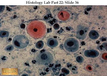
As the estrous cycle progresses, the immature parabasals are replaced by more mature cells: the intermediate cells (intermediates) (generic image from ThaiLabOnline for the moment),


the superficial cells (superficials) (generic image from bphealthcare.com for the moment)


and the anucleate squamous cells (anucleates). These are rarely seen anymore in humans, except in cases of infectious inflammation, or the wearing of pessaries, which deserves its own topic. I couldn't find any examples on the Web for that reason, so will illustrate anucleates when my graphics are working again.

All of the attributes introduced for the parabasals undergo the following regular changes as they proceed from parabasals to intermediates to superficials to anucleates:


  • their round or oval shape becomes less round and more polygonal or irregularly shaped;

  • their large nuclei become smaller and smaller, in some cases dying or disappearing altogether, so their high nucleus to cytoplasm ratio becomes progressively less;

  • their thick cytoplasm becomes thinner;

  • as their cytoplasm becomes thinner, the dark stain (high saturation) of parabasals is replaced by lighter and less intense color in the more mature types of cells.


So we can see a progression of attributes as the cells move through the estrous cycle.

Both parabasals and superficials have distinct attributes as previously described. Intermediates are all those cells that fall in between. Once a cell loses its round ovoid shape (increasing the cytoplasm-to-nuclear ratio), it should be classified intermediate. Once the nucleus degrades/compresses, thus becoming pyknotic, it should be classified superficial.

Superficials and intermediates are easily distinguished from anucleates because of the absence of nuclei in anucleates.

Distinguishing between intermediates and superficials is the most subtle distinction to learn to make, since the primary difference between them is the pyknotic nucleus of the superficial. Since the nucleus is already degenerating in the intermediate, learning to determine when the nucleus is actually pyknotic (as opposed to simply becoming pyknotic) is an important skill to master.

Up to this point, all of the examples of cell types have been in isolation. Vaginal cytology slides, however, typically contain a mixture of cell types. The proportion of each cell type is the maturation index, and the value of the maturation index---i.e., what type(s) of cells are in the majority---indicates what stage of estrus the animal is in.


Read more!

Cell classification: Parabasal cells of the vaginal epithelium

(until I get my graphics problem resolved, I'm just going to link to some generic images on the web for the sake of illustrating the point.)

Cell comparison 1: Immature vs. more mature cells

Parabasal cells (parabasals) are the youngest cells in the estrous (maturation) cycle. The distinctive attributes of the parabasal cell are:


  • their shape is round or oval;

  • they have large nuclei in relation to their cytoplasm (high nucleus-to-cytoplasm ratio);

  • their cytoplasm is thick;

  • they generally take a dark stain (their color is highly saturated).


The examples of parabasals in this image from Loyola University Chicago demonstrate all of these attributes.


They are rounder than the more mature cells which will be shown in the upcoming posts. Their nucleus-to-cytoplasm ratio is greater, and their color is darker than in the examples of mature cells. In these characteristics, the human parabasal resembles that of other mammals, such as the dog and the cat.


Read more!

Saturday, August 06, 2005

Overview of mammalian vaginal anatomy and the estrous cycle

(technical problems with the illustrations for this post--sorry! I should have it fixed by Sunday morning. UPDATE: sometime Monday, instead--the DSL here and the firewall are fighting, and I need to get some real help, which means Monday.)

1. Cross-sectional view of vagina

These photographs show the physical cross-section of the vagina at two different stages of the estrous cycle. At the top of the photographs are the more superficial layers; at the bottom of the photographs are the deeper layers.

(generic image from Wisconsin here, until I can get my images to work)


Distinct kinds of cells that appear quite differently can be seen within these layers. These different kinds of cells will be discussed in more detail in these posts

The first photograph shows the vagina with its epithelial cell lining during the proestrus stage. The second photograph shows the vagina with its epithelial cell lining during the estrus stage. Note the layer of cornified (keratinized) cells which lines the vagina during the estrus stage.

(15)

Figure: cross-sectional view of vagina

2. Description of the estrous cycle

This diagram is a schematic after Schutte 1967, representing the same view of the physical vaginal cross-section shown in the previous photographs. Under the influence of estrogens, the cells proceed from the proliferation stage to the differentiation stage to the exfoliation stage. The cell types which are representative of each stage are magnified in projection, and shown along with their names and descriptions.

The correlation between the types of cells and the physical vaginal layer is shown on the left (deep <--> superficial), and the correlation between the types of cells and cell age is shown on the right (immature <--> mature).

Note that there are significant differences between Schutte's classification scheme (indicated in black) and the one we will follow (indicated in red). We distinguish anucleate squamous cells from superficials (with pyknotic nuclei), both of which Schutte classifies as superficials. In turn, he distinguished between large intermediate cells and small intermediate cells, but we do not---we put all intermediate cells in the same category.

(17)

Figure: Description of the estrous cycle

3. Estrous cycle across time

This schematic (after Schutte 1967) shows temporal changes in cell type across the estrous cycle. For clarity of presentation, only the proestrus, estrus, and metestrus stages are shown. The cells are shown in pink and blue, because color is not definitive for cell classification---the same type of cell can appear in different colors, depending on pH. Shape is a much more reliable criterion classification than is color.

The quantity of parabasal cells at the beginning of the proestrus stage yields to a mix of more intermediate cells, which in turn are joined by superficial cells and anucleate squamous cells as the stage progresses into estrus. As the estrus stage turns into the metestrus stage, the number of intermediates, superficials, and anucleate squamous cells decreases, and the number of parabasal cells increases again.

This schematic also shows the hormonal (estrogen) levels through the different stages. Estrogen peaks during the estrus stage, and then falls off sharply. Additionally, leukocytes (white blood cells) proliferate during the metestrus stage, and then decline during the proestrus and estrus stages.

(19)

Figure (done)


Read more!

Pap smears

Before we get into the details of the cells of the mammalian reproductive cycle, let's review what our instrument, the Pap smear, is.

The Pap smear is a diagnostic test named after George Papanicolaou, a Greek physician and zoologist who developed the test in the early 20th century. Although nowadays the test is best known as a screening tool for cervical cancer, its origins actually lie in Papanicolaou's observation of regular vaginal cytological changes over the course of the estrous cycle of the guinea pig. He repeated his observations in a variety of species, for which the pattern of changes was validated, and decided to apply it to humans, using, among others, his wife (who was obviously a very good sport!) as a subject. He was able to demonstrate that the varying proportions of the different types of vaginal epithelial cells in smears on the slides were a reliable indicator of the reproductive stage the subject was in at the time the slide was taken. This proportion came to be known as the maturation index (MI), and is still used as an indicator of reproductive stage in humans and other species.

The technique for the Pap smear is essentially the same one Papanicolaou used in his studies on guinea pigs. Its use as a cancer diagnostic was actually a by-product of this original goal of investigating reproductive cycles. When Papanicolaou began testing his technique on humans, one of the women involved in the tests happened to have cervical cancer, and his observations of her particular cytology in the vaginal smear led to its use as a cancer-screening tool.


Read more!

So much death lately

Part of the reason I haven't been blogging and have been so out of sorts.

So as an antidote, I think I will blog about life, specifically the reproductive biology aspect. Part of my research involves creating comparative anatomy information systems, and one of the reasons that we know so many things about human biology from mammals, and vice versa, is that as mammals, we all share some underlying reproductive similarities, along with specific differences. My interest is in gathering common information about mammalian reproduction, and implementing it in a system that can be used to promote reproduction in endangered species.

So without any further ado, it's time for Pap smear blogging!

"We shan't save all we should like to, but we shall save a great deal more than if we had never tried." - Sir Peter Scott, founder of the World Wildlife Fund


Read more!

It ain't pretty

But it is finished (and it was loads of fun, too!). The reason I got such a great deal on my room in Philly is that it involves a modicum of house, pet, and yard work--and today I got the nerve to get on the riding mower for the first time and tackle the lawn.

It was a blast, zipping along in 3rd gear playing Hank Hill, although the yard looks like the horticultural equivalent of an amateur haircut. But it is serviceable, and I learned some lessons for next time.

It was more Sorcerer's Apprentice than Martha Stewart, though--after an initial success, I tackled a hill that, in retrospect, was probably too advanced. Despite what I've heard, I think it is possible to tip over a riding mower--not that I did, but the going was a little dicey on that hill. But no disasters, the lawn overgrowth from our recent rains is gone, and I'm that special kind of tired that is infused with a sense of accomplishment.

Not having a car, I'm tempted to reënact The Straight Story and drive into Philly this weekend, but I probably better not.


Read more!

Ya know...

If you're going to drive around with a vanity plate "Chozin" and a license frame asserting that "In case of rapture, this car will be unmanned", you probably shouldn't tempt The Lord Thy God by running a red light in morning rush hour.


Read more!

Belated post: Urban soundscape

(this was composed when James Doohan passed away; it is being posted very much later.)

Keith Olbermann was a little off his game today (though it must be said that even when he swings and misses, he's still better than a lot of anchors at the top of their game)--first, he said that James Doohan was the first Star Trek actor to pass away (if by that we stipulate that we mean major recurring character, that sad distinction belongs to Bones) and second, he said that the phenomenon of birds imitating cell phones is so far limited to Germany. Unfortunately, he is talking out of his cloaca on that one.

I beg to differ that the phenomenon is limited to Germany. The parrots we house-sit for all have their own sounds, and sometimes, they'll begin riffing off each other, picking up and developing what we refer to as "Urban Soundscape". From the aviary come the distinct sounds of car alarm, truck backing up, and--yes--cell phone ringing.

Maybe we should record that for Iain's Animal Sounds program, though parrots are already represented.


Read more!

Goodbye good and faithful friend

Godspeed, Top Dorothy. You were a good hamster, and I will miss you.


Read more!

A very belated goodbye: RIP James Doohan

I had an opportunity to arrange to be where he was "just by chance" a couple of years ago--but the way I got that information involved someone else inadvertently committing something that later would become a HIPAA violation--and so I couldn't justify carrying it out to myself. I always hoped that instead someday I would get a legitimate chance to meet him, and sadly, that is never to be.

Thank you for years of entertainment since I was a child, James Doohan--you are an actor and a gentleman.


Read more!

These cats will be the death of me

Between one cat's intermittent presents of dead mice, and the other's health problems, there have been some exciting moments cat-sitting this last couple of weeks.

One morning last week, as I was leaving for work, I heard a loud thumping sound upstairs. I thought it must be a raccoon who'd gotten into the attic to get out of the heat. Climbing the stairs, I was looking forward to finding out just what exactly I would do about a wild animal in the attic, as I had no preconceptions on that point--but when I got to the top of the stairs, it was clear that the sound was not coming from the attic. The cat was having a huge convulsion, flopping and thrashing around on the floor.

This was really scary to watch; I didn't know if she was dying, but from the violence of the seizure, it would not have surprised me. I was scared to touch her, because I thought I might inadvertently injure her, but when she started to flop over the balcony, I did grab her by the nape of the neck and gently pull her back. Otherwise, though, until the seizure stopped, I didn't know what to do.

Finally, it stopped, and Cat looked pretty disoriented, so I thought I should get her to the vet right away. Not having a car here, though, that's non-trivial, so I checked the bus schedule and found a bus would be along in fairly short order. We took the bus up to the cat's regular vet, but were turned away because I couldn't prove that I was supposed to be taking care of the cat, and because they don't take walk-ins, only appointments.

Fortunately, there was another vet a bit up the road, and rather than make the cat wait for an hour in the hot sun for another bus, I started walking uphill. Now, Philadelphia has been going through several heat waves this summer, and this was one of the worst days yet. So I'm walking up the hill, in the heat, no transportation, not knowing what to do about the cat if this vet won't see her, no extra money to pay for the bill as I haven't gotten my first paycheck yet, and the whole thing starts getting overwhelming, and I burst into tears. I got out the cell phone and called my best friend back in Seattle, "Dorothy, I want to come HOME!". As I walked along the road with the cat, she talked me down off the water tower with some sanity about how well things were really going in Philadelphia (not in the moment, but in the long run), and by the time I got to the vet, I was calm enough again to explain the situation.

The vet checked Cat out; she couldn't find anything wrong except a certain unsteadiness, but recommended further tests. I told her I'd get back to her on that after communicating with the owner, but as she's abroad, sometimes it's a few days before I can get an answer.

I was walking back home when the carrier broke open, and the cat landed on the sidewalk. I had visions of going to all that trouble to save her life, only to lose her under the wheels of a car. Fortunately, she was so stunned by her successful escape that she just stood there looking confused for a moment (my Daphne, on the other hand, would have been running before she hit the ground), and that was all I needed to grab her, and stuff her back into the carrier, which I then carried home in a death grip to prevent another escape. I could just feel the cortisol stress hormone coursing through my veins, but we made it home without incident.

With transportation and everything, I ended up taking the whole day off work, but my coworkers were extremely kind and understanding about it, and reassured me that of course I had to take care of the cat. By the end of the day, I was feeling better, and emailed the cat's owner what had happened.

A few days later, I got an email back from the owner: "Sorry, I forgot to tell you--she does that about once a month. I just wrap her in a towel until it passes.".

Someday, this is going to be a really funny chapter to look back on in my mémoirs--but I can tell, I'm still not quite there just yet.


Read more!

Dead mouse cleaning

You know, if you'd asked me before I took this cat-sitting gig, what my preference in cleaning up dead mice from the living room floor would be, in the lack of any experience I'd have gone with guessing fresh-killed over indefinitely-long-dead in provenance.

Just goes to show you how naïve I was--I now have enough data points to disprove that hypothesis. I had totally underestimated how helpful rigor mortis is in picking up a dead rodent with minimum contact. A freshly-killed, still warm, mouse is floppy and squishy and hard to manage through the layers of plastic, while a mouse in rigor can be easily picked up with two fingers and a wad of plastic, and then simply flipped into the garbage bag.

Bet you won't find THAT tip in Martha Stewart, eh?

And the cat has now earned more scrutiny at the border as a result--he used to be allowed to pass in and out freely, but now I keep the screen door closed. If he wants back in, he's got to show me an empty mouth and paws first. Sorry to be a hard-ass, Cat, but you started it with that streak of dead mice.


Read more!