A leg to stand on
While cleaning up my hard disk post-one trip and pre-another, I found the following little presentation from my comp anat class of a couple of years ago. I had fun doing it and presenting my results, so I’ll share it here as well.
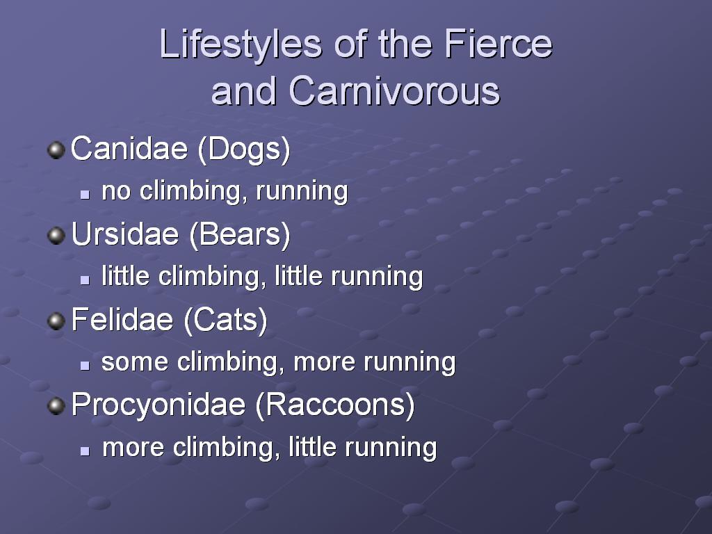
This was the independent project option for the course. My teacher Karen suggested a hypothesis—that the angle of the femur head in relation to the shaft was correlated with the running vs. climbing lifestyle in carnivores—and so I investigated that hypothesis for that part of my final grade.
The presentation proceeded in the standard format of a scientific article.

First, we talked about the femur, its structure and function, reviewing some of the literature on femurs and carnivores. Then we introduced the hypothesis.

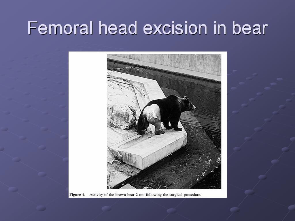
While I love theoretical mathematics and biology, it's important to remember that these ideas we are discussing have real-world consequences, as well. While the hypothesis is linked to a theoretical understanding of evolution, it is possible that the better understanding that comes out of this research can someday be translated into better clinical care as well. This slide shows a black bear recovering well after the vets in a European zoo cut out the head and neck of her femur.
PubMed reference:
Witz M, Lepage OM, Lambert C, Couillerot D, Fau D, Rachail M, Troncy E. J Zoo Wildl Med. 2001 Dec;32(4):494-9. Brown bear (Ursus arctos arctos) femoral head and neck excision. Surgery/Anesthesiology Unit, Small Animal Department, École Nationale Vétérinaire de Lyon, Marcy-L'Étoile F-69280, France.
A 30-yr-old untamed European female brown bear (Ursus arctos arctos) with a craniodorsal luxation of the right femoral head and bilateral degenerative joint disease of the coxofemoral joint had a femoral head and neck excision following unsatisfactory conservative medical therapy. The bear was injected with zolazepam-tiletamine, and anesthesia was induced with i.v. thiopental and maintained with isoflurane in oxygen via endotracheal tube. A lumbosacral epidural injection of medetomidine-bupivacaine provided additional analgesia. Slight initial cardiorespiratory depression was counteracted with fluid and inotropic drug administration and ventilatory assistance. The bear's gluteal muscle anatomy differs from that of the dog. Recovery was uneventful. The bear was confined indoors for 6 wk and was able to ambulate normally within 6 mo.
Notice the part of the abstract that I have emphasized in bold--if the bear's muscle and bone anatomy differs from that of the dog, then knowing that fact helps the veterinarian take better care of both bears and dogs. Perhaps having a bear live in an environment suited to dogs caused undue wear on those structures, for example, and can be prevented or better treated nonsurgically by understanding what enviroment the bear needs, as opposed to other species. Or, if surgery is necessary, knowing what variations you find before you go in can cut down on the time you keep the animal under anesthesia while exploring its anatomy. Understanding the evolutionary theory behind that improved clinical care places the knowledge in context, and helps develop the next hypotheses that will be tested to improve both our understanding and our care for these animals.
This slide shows another case study in a wild carnivore, this time a red panda.
PubMed reference:
Delclaux M, Talavera C, Lopez M, Sanchez JM, Garcia MI. J Zoo Wildl Med. 2002 Sep;33(3):283-5. Avascular necrosis of the femoral heads in a red panda (Ailurus fulgens fulgens): possible Legg-Calve-Perthes disease. Zoo-Aquarium de Madrid, Madrid, Spain.
A 17-mo-old captive-born female red panda (Ailurus fulgens fulgens) presented with a sudden onset of lameness in its left hind leg was diagnosed radiographically as having possible severe, bilateral Legg-Calve-Perthes disease with fracture of the great trochanter of the left femur. Surgical repair of the fracture was performed using pins and a tension band wire through a lateral approach to the hip. This is the first case reported at Madrid Zoo-Aquarium, where 63 individuals have been bred over 15 yr.
The same points I made about clinical relevance for the black bear are true here, so I won't repeat myself, but let's look a little closer at the X-ray to see what's going on. First, we'll orient ourselves: the caption says it is a "ventrodorsal" image, so that means that we are looking at it starting from the abdomen and going toward the back. So we are looking at the panda's belly, rather than looking at its back.
Ok, so that orients us front-back; now let's figure out where up and down are on this image. The panda is anesthetized for the X-ray, so it is totally relaxed and asleep. Think about how a similar animal--a cat, for example, stretches out its hind legs when it sleeps. The legs are stretched out behind it, so since the legs are stretched out in the "up" direction in the image, "up" here must mean "behind", and "down" is "front". Looking at the vertebrae reinforces this, as the vertebrae at the bottom of the image are larger (backbone) than those at the very top (tail). So now we know we are looking at the panda's belly, and the panda is upside-down, relative to us.
We know 2 of our 3 dimensions, so now we can figure out the third--if we are looking at the panda's belly, and the panda is upside-down in the image, then we can rotate the panda image to face the same way as we are, determine left and right, and then rotate it back, seeing what happens to left and right as we do so. To get the panda to face the same way we are, first we have to flip the image from top to bottom. That means the image's original left becomes right, and original right becomes left.
Now the panda is right-side-up and facing us ("ventro-" [belly] in "ventrodorsal" , so we have to rotate it around to face the same way as we do to determine its right and left. So original left, which became right above, becomes left again, and original right, which became left above, becomes right again.
So the left and right of the anatomical position of the panda matches the left and right of the image--now we can see what they say about the image, and we are oriented correctly to look for what they want to point out.
The radiograph's caption says that the right femoral head is deformed, and that the left femoral head and neck are missing. To be honest, I am not so good at reading X-rays that I can tell the difference myself--for our purposes, let's just remember that they should be more or less bilaterally symmetrical. Looking at the X-ray, on the other hand, we can see that there is a great difference from one side to the other on where the hind legs join the pelvis (the head of the femur)--they do not look very similar at all. This is another case study of how animals can develop pathologies of these structures, and how understanding the reasons in the species-specific variation of these structures can help us to not only forward the science )as useful a goal as that is in itself_, but can also translate to better preclinical and clinical care for the animals involved.

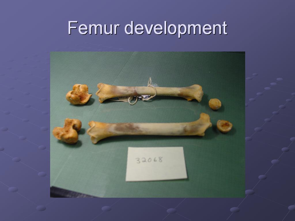
The parts of the adult femur. When I was developing the plan for this project and before I first began examining femurs, I wasn’t sure that I would ever learn to tell a femur from a humerus, but that actually turned out to be trivial, once Karen went over the parts with me one time. The head of the femur is unique, and once you have learned to recognize it, you will never confuse a femur with a humerus again.
Notice the mushroom-shaped structure at the top left of the femur on the left, and on the top right of the femur on the right. That is the head of the femur, and the structure we are primarily looking at. It is the structure we saw on one side of the panda X-ray, but not on the other side.
The rest of the femur (minus the head) is what we are referring to as the shaft of the femur. You see how the head forms kind of a 45-degree angle to the shaft. The variation in that angle (of where the head attaches to the shaft) is the variable that we are most interested in.

What did trick me in the beginning, however, were these bones. Often, a box of animal bones would have no obvious femurs, but would have two sets of these. Turns out, these are femurs which are still developing—femurs from a juvenile animal (in this case, a raccoon).
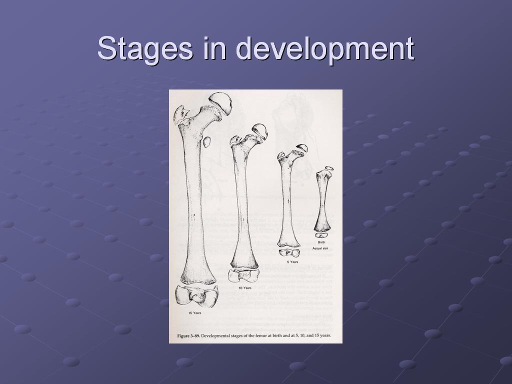
This diagram shows the ossification or bone formation and hardening patterns of the femur. Notice the separate parts that we saw in the raccoon femur in the previous slide are shown here in the way in which they grow together and solidify.

Not quite sure why they chose to show these in the counter-intuitive right-to-left order, but from right to left, this progression shows how the femur develops from youngest to more mature, the leftmost image showing all the adult parts right before they are finally fused.
We used a sample of the animal bones in the collection of the Burke Museum. We chose four groups to take representative samples from for comparison. Additionally, we ordered them by the degree of climbing and running they normally do.
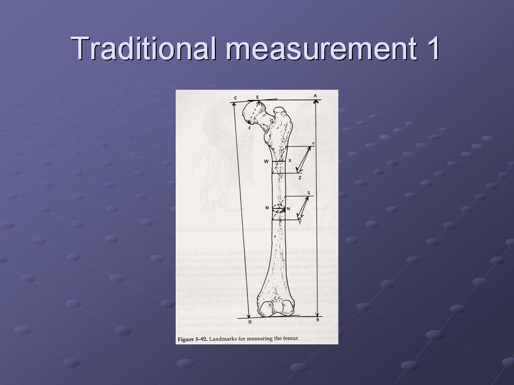
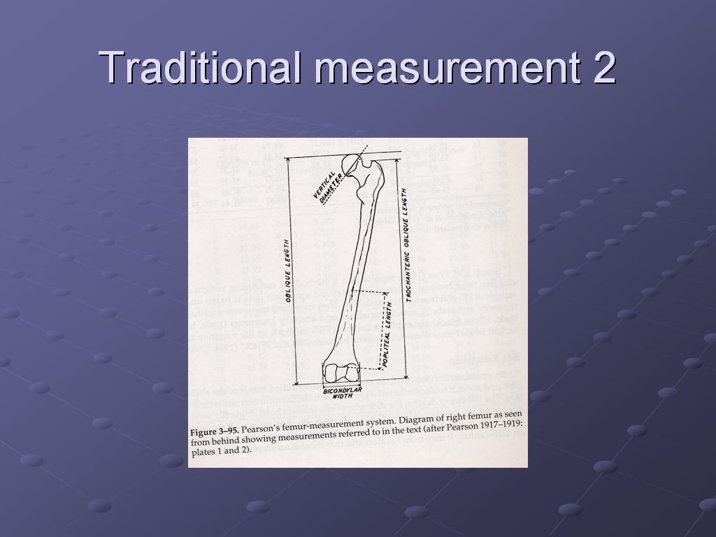
These two slides show traditional ways of taking femur measurements. Because of the time constraints on the class project, and the lack of access to precision instruments, I came up with a method of simultaneously measuring and recording those measurements to save time. While this could generate data of a quality acceptable for preliminary results of a pilot project which paved the way for a larger project, for the larger project itself, it would be extremely poor form to try to both test the hypothesis and to validate the instrument used to test the hypothesis at the same time.
Karen’s project suggestion, stated as a hypothesis.

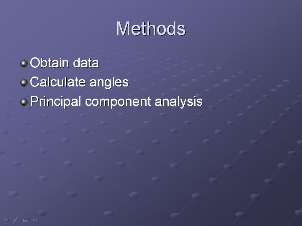
My methods were to 1) obtain data from femur specimens of the Burke Museum's mammal (specifically the carnivores) collection; 2) calculate the head-neck angles (using an instrument I developed), and 3) use principal components analysis (explained below) to cluster the data by species, and see if the expected trend was present.
This slide shows my setup. To consistently measure angles, I built a box in 3D to hold the femurs while I photographed them, and later gathered the desired measurements from the photos. I printed sheets with 1-mm squares on them to use as a grid, laminated the sheets for stability, and then anchored the laminated sheets inside a plastic box to hold them steady and to provide an "origin" coordinate at which to place the bone.

Here are the results, post-principal components analysis (PCA). PCA is a way of simplifying the data--if you think of the linear algebraic matrix representing the object as occupying n-dimensional space, that representation and it manipulations may be so computationally-complex as to make it unmanageable. PCA simplifies the problem by transforming the representation into fewer, more manageable dimensions, chosen so that the first dimension in that space represents the parameter that accounts for the most variation in the data.
Species turned out, as expected, to be the first PCA component for head-neck angle. The website I found to run the analysis happens to be a French-language site, which explains why the chart is in French.
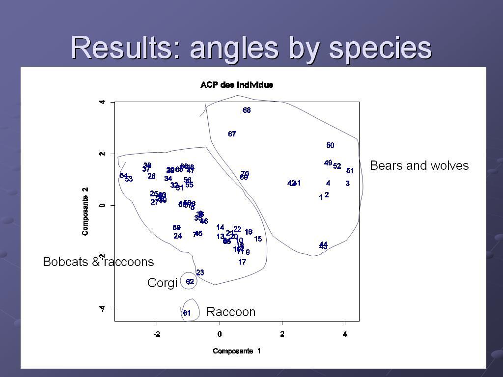

By contrast, another measurement I took--femur curvature--did not follow the same trend as head-neck angle.

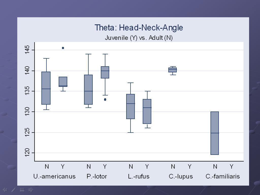
So the data very tentatively bears out my hypothesis. There was no significant variation in head-neck angle by age or sex (which I was able to obtain from the museum database), but the head-neck angle does vary as expected to correlate with the proportions of lifestyle devoted to running vs. climbing. This is a heavily-caveated conclusion, subject to the limitations described below.
Notice that there are two outliers, a Corgi and a raccoon. The Corgi was mislabeled "wolf" on the sample box, and generated some very unexpected "wolf" data; it was subsequently determined from the database that it was actually a Corgi. The distorted legs of the Corgi would be expected to make it an outlier among canids in any case. In keeping with the "As randomized, so analyzed" principle of intention-to-treat analysis, I left it in, even though it is clear that it deviated significantly from the "wolf" criteria.
This experiment had a very small sample size, a problem further exacerbated by dropping off several really large bears (because of instrument limitations), and young male raccoons (explained in more detail below). If this were more than a student class project, these limitations would have to be addressed head-on in order to argue that the data from this project was a sufficient pilot to justify a larger study.
Possible skewing of data, and an emergent hypothesis: Many of my young raccoons selected had to be excluded from this study because I had no consistent way of measuring femurs that were in pieces because they were not fully ossified. Almost all of this population were male, skewing my data not only for raccoons, but also for male raccoons. A question that comes out of this artifact is: why is the Burke Museum's sample collection skewed to collect young male raccoons? Do young male raccoons experience an early death by car (roadkill collections) at a greater rate than female raccoons because of hormonal or other influences at adolescence (in other words, do young male raccoons take greater risks in search of a mate than do either female raccoons or males of other species)?
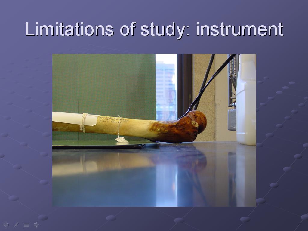
Size of instrument: Clearly, the box was not big enough to handle the largest samples. So the very largest bears were necessarily excluded from the study, and such systematic exclusion runs a real risk of skewing the data.
Parallax was a problem with my instrument and the camera I was using. For huge projects, such as architecture, special cameras are used to compensate for the optic distortion that occurs; I thought that the optic distortion at the small size of this project was not likely to be an issue. Yet, this slide shows how even at this size, the same femur in the same position looks much closer to the end of the box in the upper picture than in the lower. Clearly, I should have taken optics into account in gathering data for this project, which makes the preliminary data more suspect.

Another potential issue--my instrument did not stabilize the femurs, and so it is possible that enough variation in placement occurred to make my measurements suspect on this parameter as well. This slide shows how femurs are stabilized for medical imaging; perhaps a loose femur needs even more normalized and stable placement in order to obtain reliable measurements.

I conclude that in this prliminary data, the correlation between running/climbing lifestyles and femur head-neck angle was borne out. This conclusion is, however, tentative, and needs further replication in order to be sure.

I appreciate all the help that was provided to me--it was a fun project, and I learned a lot from it!

posted by thalarctos @ 6:59 PM
0 comments
![]()

0 Comments:
Post a Comment
<< Home