Sunday, October 30, 2005

Presentation of my dissertation work

This week, I presented my dissertation work at the American Medical Informatics Association conference in Washington, DC. The entire panel (4 papers) on ontology mapping took place, aptly enough, in the Hilton's Map Room.

Ever wondered what happens when two papers on a panel have the same title? I inadvertently found out when Olivier Bodenreider and I independently gave the same title to our papers on mapping mouse anatomy to human anatomy: "Of Mice and Men", and both were accepted. So the first and third papers on the panel were each entitled "Of Mice and Men", and when Mark Musen introduced the panel, he quipped that we would retire afterward for refreshments to the Steinbeck room.

My talk presented my dissertation work, which addresses a very important problem: the accurate determination and representation of anatomical correspondences across species, in this case, human and mouse.


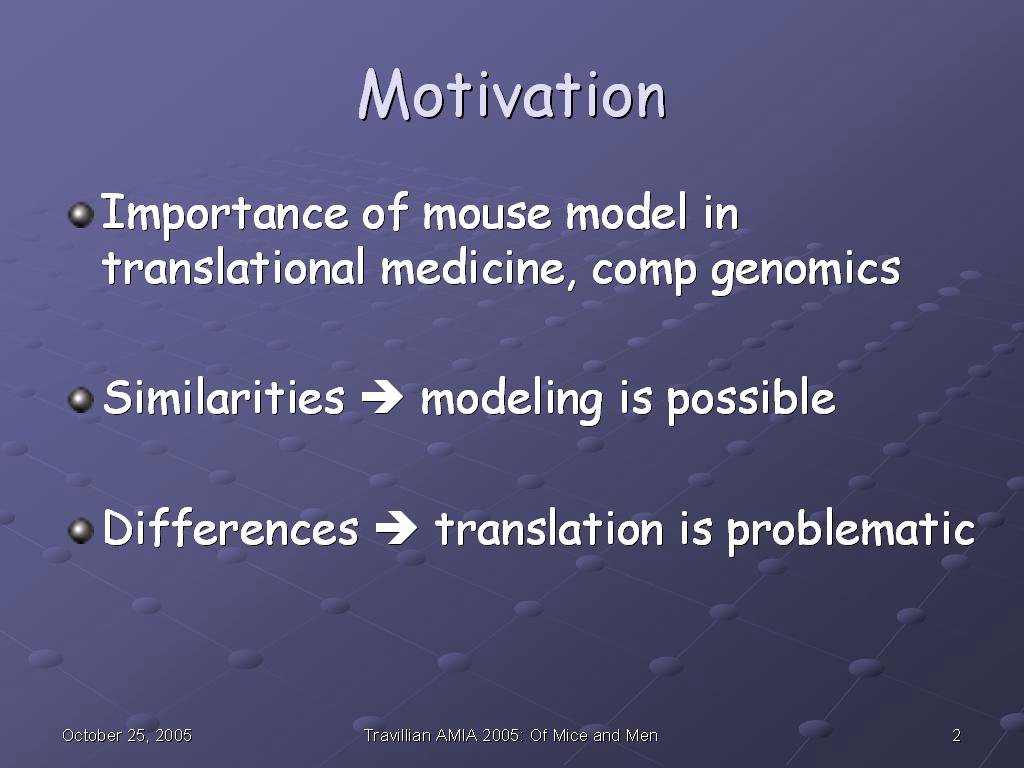
The importance of this problem has been well-documented; in fact, many of the people present in the audience had written on just that topic. Mice, as well as many other species, are used as model organisms for understanding disease in humans, and for deciphering the genome. They can serve this purpose because of the similarities between them and us, and it is these similarities which make comparative medicine possible. At the same time, the differences between us create issues in the interpretation and translation of results in these species to humans: treatments that cure cancer in mice don't necessarily do so in people, and there are reports of cardioprotective effects of Vioxx in mice, rats, and marmosets, which did not predict the harmful effects that drug would have in humans, just to name two examples. The effects of these similarities and differences emphasize the importance of sound and complete modeling of these structures across species, in order to support correct reasoning about the implications.


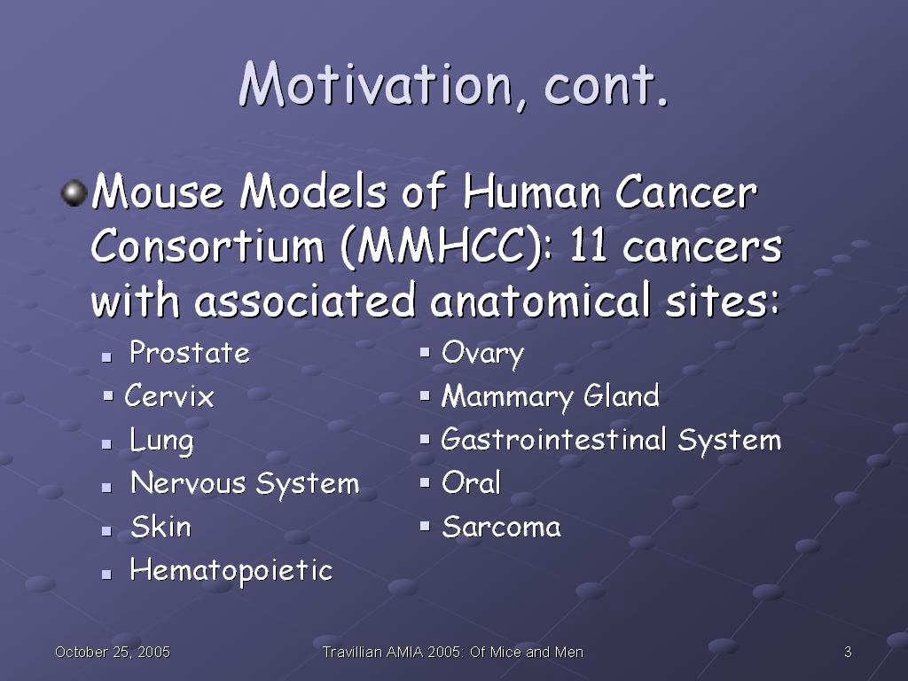
Because of the importance of the mouse as a model for human medicine and genomics, the Mouse Models of Human Cancer Consortium has identified and prioritized 11 types of cancer to consolidate murine information on. The cancers, listed on this slide, are each associated with specific anatomical sites, which is where our anatomy ontology and information system come in.


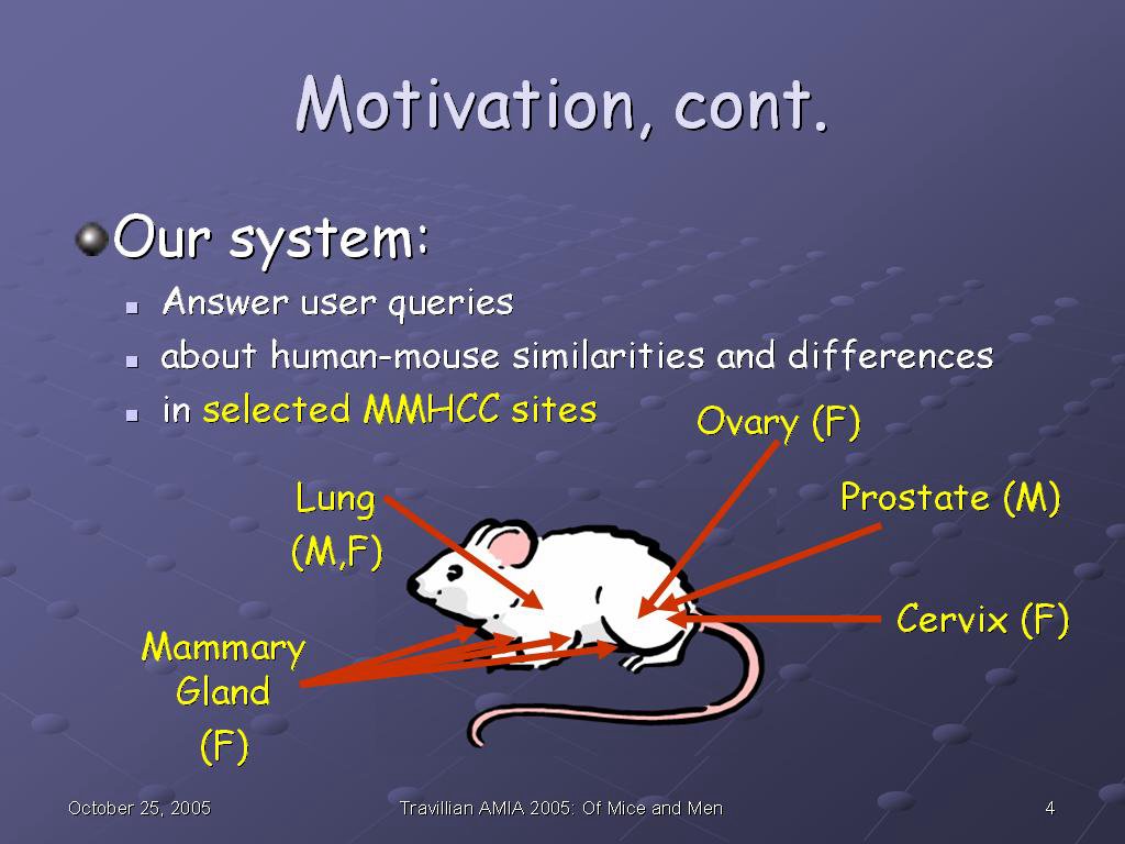
Our system is designed to answer user queries about the similarities and differences in human and mouse anatomy in selected sites taken from those identified from the MMHCC. The sites identified are the ovary, cervix, and mammary gland of the female mouse, the prostate of the male mouse, and the lung, shared by males and females.


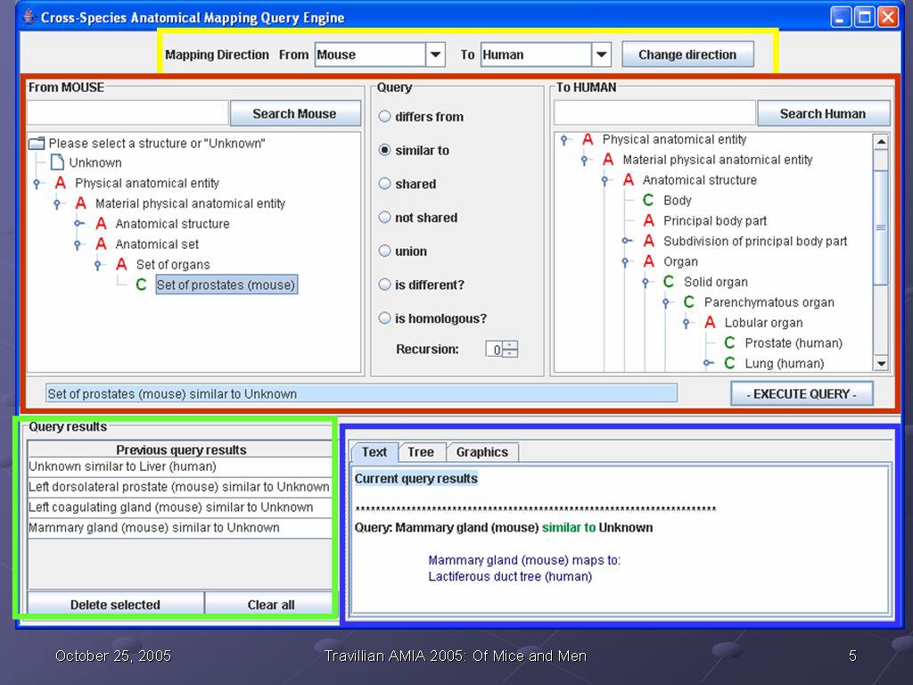
This is a screen shot of our application. There are four main areas, which are outlined on the screen.

The yellow box outlines the query direction. Users can choose to ask queries about the human compared to the mouse, or about the mouse compared to the human, as they prefer. In this way, the interface conforms to the user preference. However, as outline in my earlier MS thesis, the query is bidirectional: the user can ask the question in either direction and get the same result. In other words, the queries "how does the human prostate differ from the mouse prostate?" and "how does the mouse prostate differ from the human prostate?" return the same information. The user does not need to be concerned that the order of the query has any informational significance; it is only a user interface accommodation to the user's preference.

The green box at the bottom left is where previous queries and their outcomes are stored. A session can be cumulative in the sense that information can be gathered, and that information then used in later queries. In this way, our application builds on the work done earlier in our research group on Emily, a query interface to the Foundational Model of Anatomy. Emily also stores results of previous queries for reuse; as this was a most useful feature, we incorporated it into our system.

The blue box at the bottom left is where the current query results are returned. In the eaxmple in this slide, the user has just asked what structure in the human corresponds to the mammary gland in the mouse, and the system has returned the information "Mammary gland (mouse) maps to lactiferous duct tree (human)". What is displayed in this area is always the results from the last query submitted.

The red box in the center of the screen is where a new query is formed, and will make up the bulk of this talk. It consists of a "from" hierarchy (can be either mouse or human), a "to" hierarchy (either mouse or human), and a set of possible queries in the middle that can be selected by radio buttons. The text box below is populated as the user forms her query: in this way, she 1) does not have to remember our syntax to form a query, and 2) gets immediate feedback from the text box whether the query is what she wants, in time to change it or submit it. In the example in the slide, the user has selected "Set of prostates (mouse) similar to Unknown (human)" to ask "what structure(s) in the human correspond to the set of mouse prostates?", and the query is ready to submit by clicking on the Execute Query button.


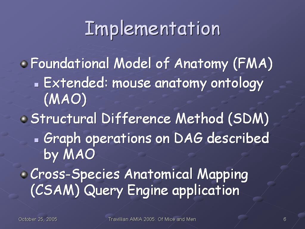
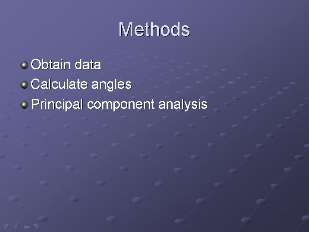
We implemented our system in the following way, described in more detail in my MS thesis and our AMIA 2003 paper: first, we used the Foundational Model of Anatomy as a template to build a partial ontology of selected mouse organs, an ontology which we shall refer to as the Mouse Anatomy Ontology (MAO).

We used the Structural Difference Method (outlined in my thesis) to perform graph operations on the directed acyclic graphs (DAGs) described by the FMA (human) and MAO (mouse).

Finally, we developed a user interface and query engine to present the result sets oobtained by the SDM to the user.


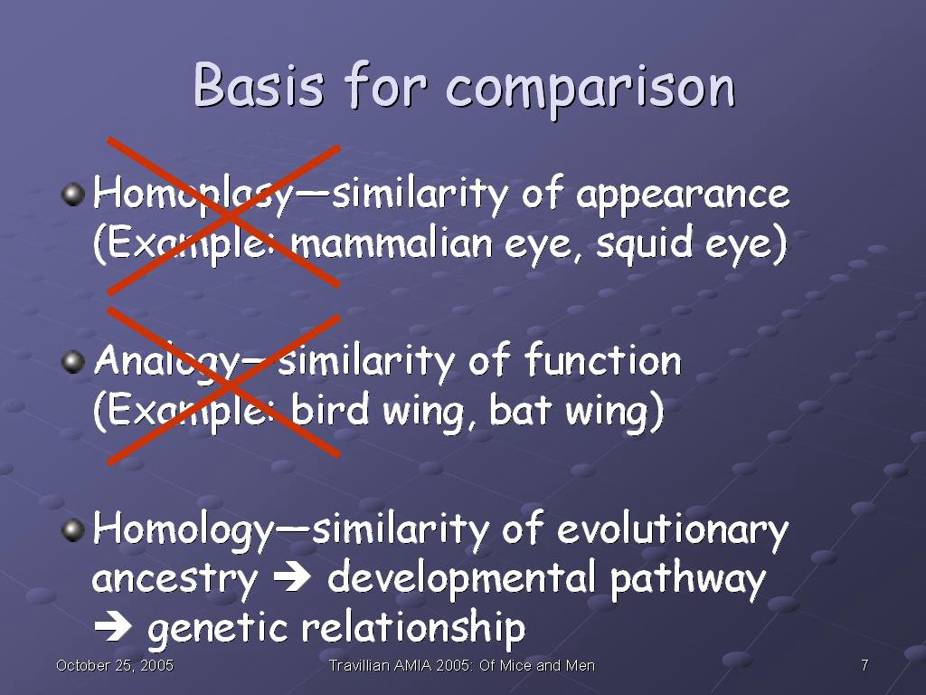
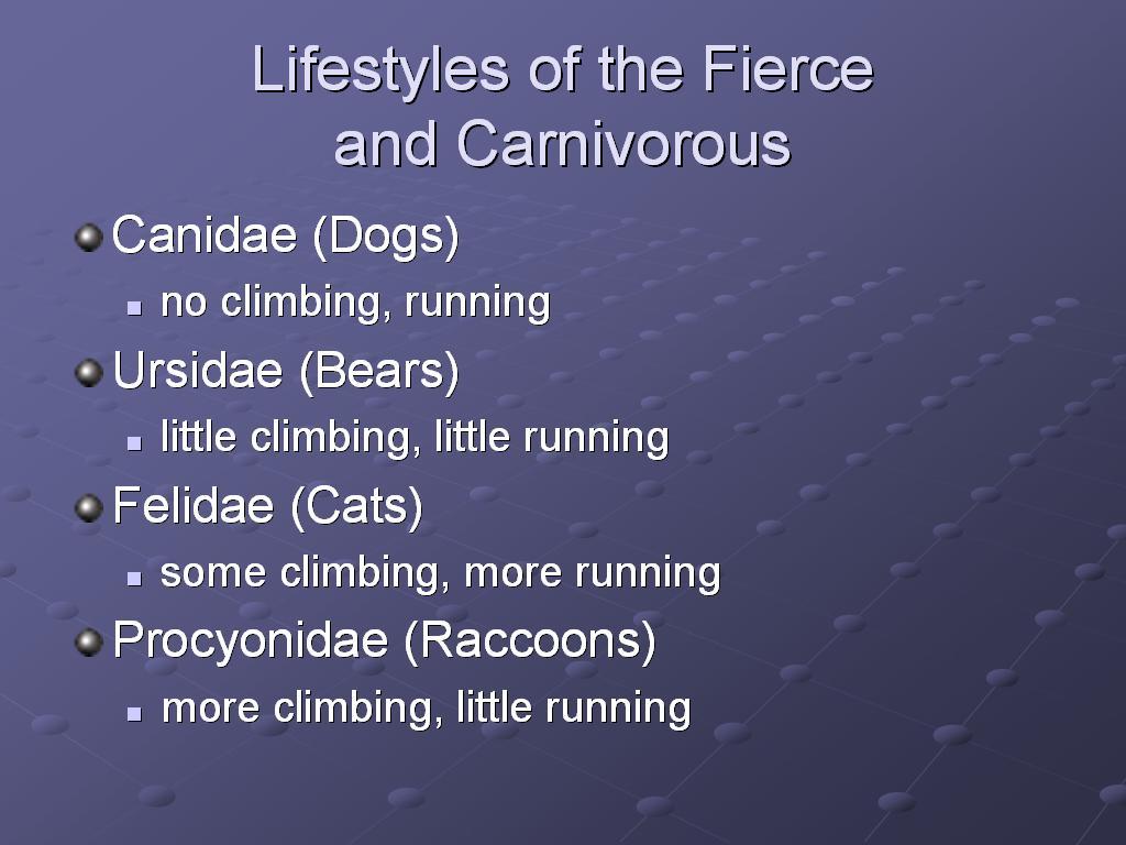
In order to determine the mappings, we had to establish what structure in one species was similar to what structure in the other species. Traditionally, when biologists and comparative anatomists have referred to "similarity", there are three different aspects that they take into account:

Homoplasy, or similarity of appearance: the mammalian eye and the squid eye appear similar to use--they "look like" eyes, which is the homoplasy. Yet structurally and developmentally, they are very different from each other. We do not model homoplasy in our model.

Analogy, or similarity of function: the bird wing and the bat wing both serve the purpose of flight, yet they evolved at different times, using different structures in the respective forelimbs involved. We do not model analogy in our model.

Homology, or similarity of evolutionary ancestry: while homologous structures may or may not exhibit homoplasy and/or analogy, it is their shared evolutionary ancestry--which we can get at via understanding of their developmental pathways, which in turn sheds light on the genetic relationships involved--that we concern ourselves with in our modeling of similarity and difference.


Because it is the homologies we are modeling, there is a natural tie-in to the Foundational Model of Anatomy ontology. As we see in the slide, by definition, Anatomical entity--a fundamental unit of the FMA--is a Material anatomical entity which has inherent 3D shape; is generated by coordinated expression of the organism's own structural genes; and its parts are spatially related to one another in patterns determined by coordinated gene expression.

The existence of the entity--coordinated by the structural genes--is what ties the FMA Anatomical Taxonomy (AT) component to homology. The spatial relationships among its parts--determined by coordinated gene expression--are what ties the FMA Anatomical Spatial Abstraction (ASA) component to homology.

A further component of the FMA, the Anatomical Transformation Abstraction (ATA), is also tied in to the developmental pathways aspect of homology; however, that is outside the scope of my dissertation, and will not be pursued here.


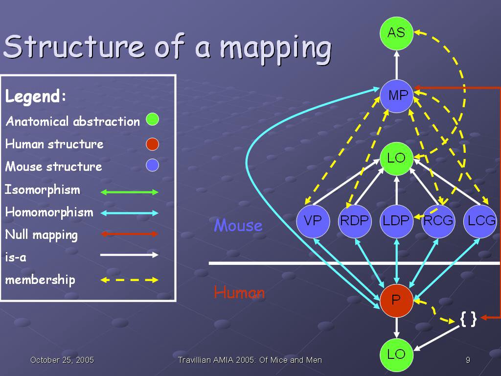
This slide shows the structure of a mapping, specifically the mapping between the human and mouse prostates. The red circle (P) represents the human prostate; the blue circles represent the mouse prostates ventral prostate (VP), right dorsolateral prostate (RDP), left dorsolateral prostate (LDP), right coagulating gland (RCG), and left coagulating gland (LCG).

The green two-headed arrow in the legend is there for the sake of completeness; there are no isomorphisms in this particular mapping. An isomorphism is a one-to-one and onto mapping between anatomical structures at a given granularity (in this case, Organ level) across species. In other words, each structure in one species at that level has one and exactly one correspondence in the other species. An example would be the heart, which is isomorphic not only at the Organ level: Heart (mouse) corresponds exactly to Heart (human), but also at the Chamber level: the human Left atrium, Left ventricle, Right atrium, and Right ventricle correspond exactly to those same structures in the mouse.

A null mapping (red bidirectional arrow) is the case when a structure exists only in one species and not at all in the other--for example, the human breast has no corresponding structure in fish models, because the breast is a structure found exclusively in mammals. So the Breast (human) maps to Null in fish.

A homomorphism (blue birdirectional arrow) is any relationship in between--it is a non-null mapping (so the structures exist in some form in both species), yet it is not one-to-one and onto (so there are structural differences of some kind which occurred during speciation). We see an example in the prostate here: the human prostate (a Lobular organ) corresponds in some way to 5 different lobular organs in the mouse, so the correspondence is 1:5 (or 5:1) rather than one-to-one and onto. It is, therefore, a homomorphism. Five blue bidirectional arrows are drawn to indicate the homomorphisms between the mouse and human organs.

There is a further homomorphism in this diagram; that between the Anatomical set comprised by the mouse organs--in other words, the mouse prostate in toto (MP)--and the human prostate in toto. Additionally, because there is no corresponding set of human prostates, there is a null mapping between the Anatomical set MP and the human (red bidirectional arrow).

The white unidirectional arrows indicate a subsumption relationship; the human prostate is-a Lobular organ, as is each organ of the mouse prostate. Further, the entire mouse prostate (MP) is-a Anatomical set. The membership (partitive) relationships (is-member and has-member) are conflated into a bidirectional dashed yellow arrow for the sake of clarity, as this diagram is already becoming quite complex, even at a level of granularity as gross as Organ.

Anatomical set is a species-independent anatomical abstraction, as is Lobular organ, indicated by the green circles. We do not map anatomical abstractions per se, as it does not make sense to say Anatomical set maps-to Lobular organ outside of any context, and as they are species-independent, there is no barrier to cross with a mapping.

What does make sense, however, is to map a structure in one species with a subsumption relationship to an anatomical abstraction to a structure in the second species with a subsumption relationship to a different anatomical structure. While there is no homomorphism directly between the anatomical abstractions themselves, the difference between the parents of the mapped structures is sufficient to indicate that an interesting anatomical transformation occurred during speciation. Therefore, although we do not map abstractions directly to other abstractions, we do keep track of subsumptions relationships of those structures which we do map.

These entities and relationships are tracked via the MAO, and similarities and differences among them are described using the SDM, and returned to the user via the CSAM interface.


Because the anatomical abstractions are species-independent, there is only one anatomical taxonomy in Protege. For example, Lobular organ is defined once--it is-a Parenchymatous organ, which in turn is-a Solid organ--and it has as children not only the Prostate (human), but also all the mouse organs. Because there is only one taxonomy, the species is appended in parentheses at the end of the structure name.


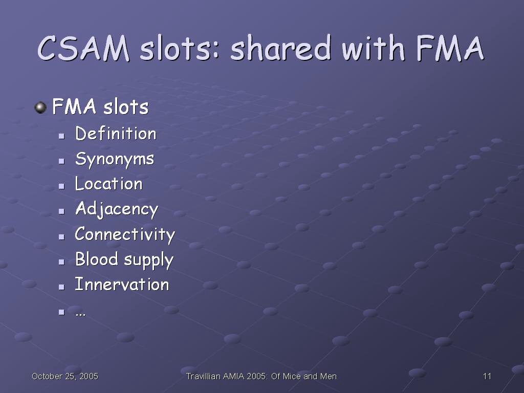
CSAM extends the FMA, and as such it has access to the anatomical information in that ontology. It get slots such as adjacency, connectivity, blood supply, and innervation, among others, from the FMA.


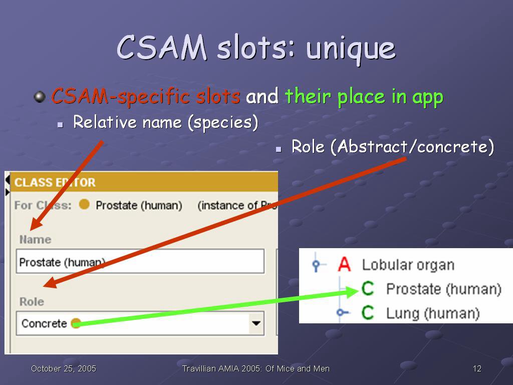
Additionally, it has slots which are unique to CSAM. One of the CSAM-specific slots, as mentioned previously, is the Relative name, where the species is appended in parentheses to the structure name. Another is the role, Abstract or Concrete, which distinguishes whether a structure can be mapped (Concrete), or--as an Anatomical abstraction--cannot, per our previous discussion

The green arrow shows how that information ends up in the CSAM interface: The red A in front of Lobular organ indicates an Anatomical abstraction, while the green C in front of Prostate (human) and Lung (human) indicates that those structures will support mapping queries.


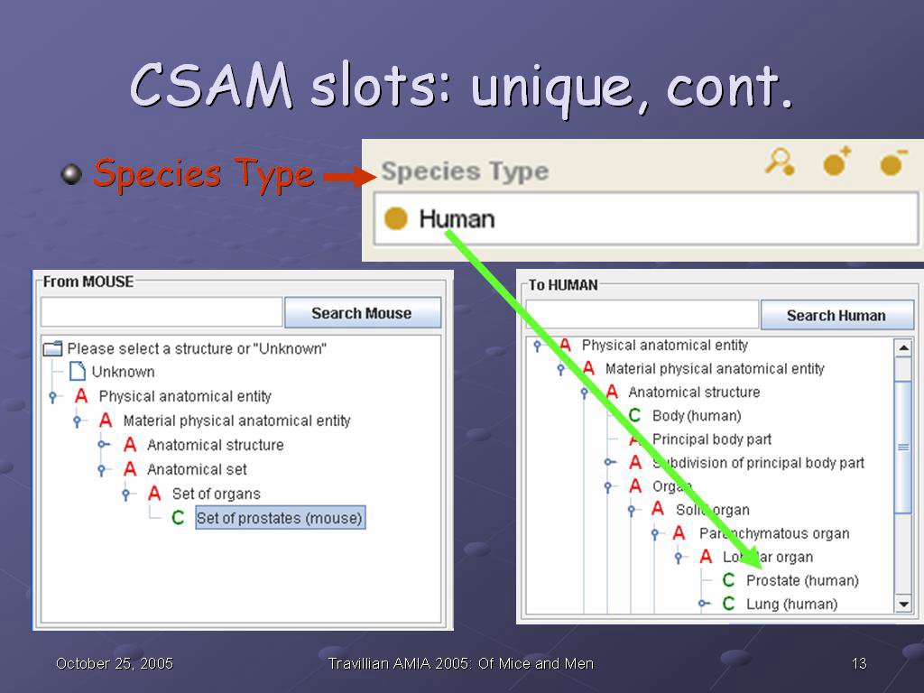
Species Type is another CSAM-specific slot, and it is used to break the one species-independent hierarchy from Protege into two species-specific ones in the CSAM interface--the From hierarchy and the To hierarchy.


The last CSAM-specific slot is Maps-to. From this slot, the similarities--correspondences (isomorphism, homomorphism, null mapping)--and the differences (union, intersection, and set complement of result set) are calculated.


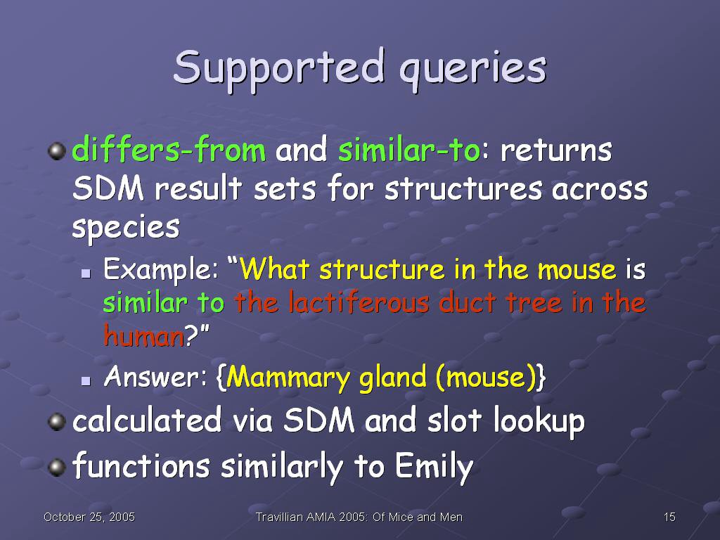
Queries currently supported include differs-from and similar-to. For example, "What structure in the mouse is similar to the lactiferous duct tree in the human?" returns the result set {Mammary gland (mouse)}. This result set is calculated via slot lookup and SDM, similarly to the way Emily functions.


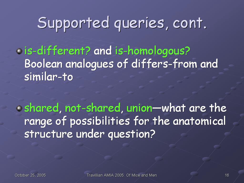
Is-different and is-homologous are the Boolean analogues of differs-from and similar-to: where the set queries ask for an unknown, the Boolean queries take two knowns and verify or dispute the proposed relationship.

Shared, not-shared, and union draw upon set and graph operations to answer more abstract queries: what structures are common across species, what structures are unique to one species or the other, and what are the range of possibilities for this structure in these species.


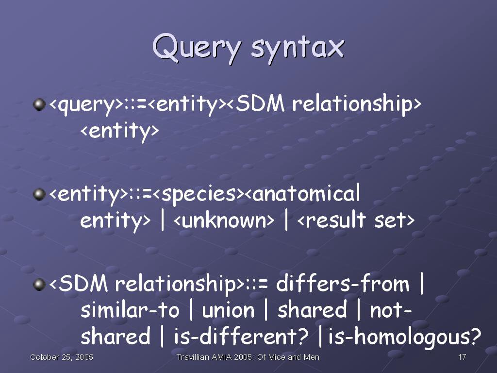
Our query syntax draws heavily from Emily: the query has three components, Subject, Relationship, and Object. Given any two in a query, CSAM can return the third component.

Like Emily, CSAM can use the result sets returned in previous queries as either the Subject or Object of further queries.

The range of allowable CSAM queries at this point comprise the ones we just reviewed.


That is the operation of the four sections of the CSAM interface: yellow box: specify directionality. Green box: store and retrieve previous query results. Blue box: display results of last submitted query (with tabs for optional tree display and display of associated graphics). Red box: specify a Subject-Relationship-Object query, enforcing correct syntax through the use of lists and radio buttons, and providing immediate feedback by translating the query into a text box.


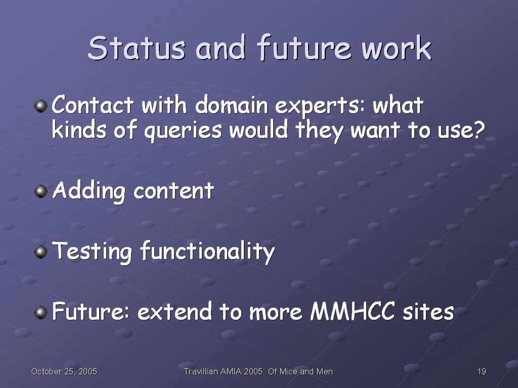
At present, I am contacting domain experts (mouse anatomy and pathology experts for the sites we have identified) to establish what kinds of queries they would want in such a system to make it fit their information needs. We are adding content in accordance with their responses. Additionally, we are testing functionality as we add that content, and we project a future project is to extend this approach to answer queries about more MMHCC sites of interest.


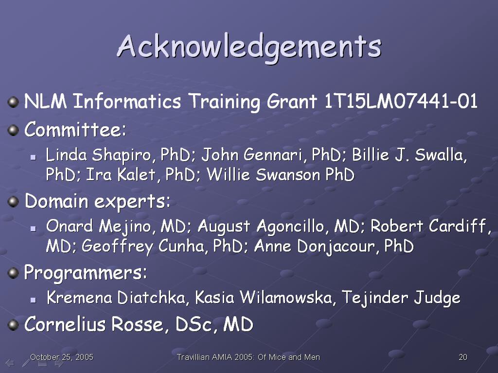
I would like to thank all who made this work possible: the NLM, whose training grant funded this work; my committee; the domain experts, of whom this represents only a small sample, as they are too numerous to name individually; the programmers who have helped me to implement my design; and my mentor, Dr. Cornelius Rosse, the original developer of the FMA.


Read more!

Saturday, October 15, 2005

Which Dromaeosaurid Are You?
brought to you by Quizilla

Utahraptor
Utahraptor - Large, yet swift, you never
let your pack down.
You play with wild abandon,
But are the most protective
of packmates.


Which Dromaeosaurid Are You?
brought to you by Quizilla


Read more!

Thursday, October 06, 2005

Did you hear the one about the ornithologist

who couldn't go to the bird conference?

He had to send his egrets.

That one remains every bit as funny as the first time I heard it (a sentiment that cuts two ways, no doubt), but the reason I bring it up is that describing the deer by the side of the road in Valley Forge reminded me of the egrets on the runway's grass strip as the plane from Albuquerque landed in Houston. Tons of metal bouncing down hard, engines immediately thrusting hard into reverse and revving to slow the plane, the loud sound of brakes being applied--all this not 20 feet away from the egrets, and, like the deer, they never even batted a feather (that sentence doesn't work: deer don't have feathers. how about:) keratinized epidermal projection. (That works both for feathers and for eyelashes :).

"Wild" animals, my asinus.


Read more!

Where the boys aren't

Valley Forge Park (yes, that one, with all the history).

Hmm, perhaps that answer needs a little unpacking to be at all understandable.

First of all, horns (mammalian, not lizard--that's totally something else) and antlers have some surface similarities, as well as fundamental differences. They each have bony cores, and are located in similar places on the front of the animal's skull. However, their compositions are different in important ways. What I always find surprising is that--despite the hardness and permanence of bone, as compared to hair, skin, and nails in the way we think of them--the permanent structure (horn) is covered with keratin (in most horned animals), or, in rhinos, is made totally of compressed keratin (basically, hair) with no bony core. On the other hand, the temporary structure, which is shed and regrown annually in deer, moose, and other cervids, is made totally of bone. Seems like it should be the other way around, given the ephemeralness of keratin in hair and claws, compared to bone, but there you are.

Male deer regrow their antlers after each mating season. When the antler is regrowing, before it is mature, it is covered with "velvet", which is a layer of skin full of blood vessels to promote the antler's growth, and which is shed when the antler is done growing. Cervid phenomenology being in its infancy, it's impossible to know for sure, but I wonder if the sensation of an antler growing is anything like I remember for permanent teeth coming in--not pain, not exactly, but exquisite sensitivity, and a desire to constantly rub my gums to make the feeling go away. If so, then the male deer have another reason (besides the mating season rise in testosterone levels) to be cranky.

Pennsylvania seems to be chock-full of deer anyway, and riding the bus through Valley Forge National Park frequently, I often see them standing near the road calmly eating grass, and paying no attention to the cars driving right by them. I hadn't thought about it until the bus driver (F.) asked me, but then I noticed he was right--we were never seeing any antlered deer.

So what proportion of antlered deer would we expect to see? We know that only sexually-mature males have antlers (in this species; in reindeer, by contrast, both males and females have them). In the absence of any specific knowledge, we can assume that the population is split roughly equally between males and females, so out of every four deer, we would expect two of them to be male.

Assuming that deer reproduce at a rate that roughly replaces every adult who dies with a younger one, let's say that the population is about equally distributed between mature deer and immature ones. So out of our two male deer, let's say that one should be mature, and the other immature. Out of our four deer, then, we have one antlered one, and three without antlers (as long as our initial assumptions about gender and age distribution are accurate).

So we should have been seeing roughly one deer with antlers for every four deers, or maybe fewer if our assumptions were wrong--but we were seeing zero antlered deer, way fewer than we could explain, even if we had been wildly off. So since our data were at such variance with our hypothesis, we started to look at possible systematic explanations for the discrepancy.

Possibly they were being deantlered, as I have heard is done in some Japanese deer parks, to protect the human visitors (turns out, no). Possibly their antlers hadn't fully come in yet (but mating season is earlier in the year, so that explanation doesn't work, either). We tried a couple of other possibilities, but nothing really made sense. Later, LL was able to supply a missing piece of the puzzle that hadn't occurred to us.

Hunters prefer deer with antlers, the bigger the better. Therefore, there is a selection pressure against antlers, and the population is skewed toward the non-antlered deer. Additionally, if the antlered deer have the least bit of sense, hiding from humans is a good thing when they want to kill you. So in addition to the antlered deer dying at a higher rate, probably the ones that aren't dying have learned to stay out of sight of people driving by on the nearby road.

Obviously, we haven't proven this in any rigorous sense, but it is highly plausible on the face of it. F. and I were very satisfied with our approach to this question and its resolution.

And, amusingly enough, now for three days in a row, we have seen a new antlered male who comes close to the road to eat grass. He must be very brave, or else very oblivious--but it is nice to see him; he is splendid with those antlers.


Read more!

You've overplayed your paw, Cat

Just how stupid do you think I am?

Oddly enough, since the cat who had the seizure continued to get sicker and sicker and finally shuffled off this mortal coil, we are going through more, not less, cat food than when we had two cats. Mystery partly solved when LL and I compared notes--turns out that after I've been feeding him and leaving for work in the morning, he goes and cries outside her door, convincing her that I forgot to feed him, and scoring a second breakfast.

Ok, I have to admit that was pretty smart. But his success led him to branch out, and now he's overreaching.

Here's a hint, Cat--for that trick to work, you need two different marks. Not having Korsakoff's syndrome, and having actually transitioned out of the concrete operations stage into the formal operational one a long time ago, I was not fooled for a minute when, after I fed you and went back in my room, you stood outside my door and cried about how Raven forgot to feed you.

Not gonna work, Cat--you're going to have to pull something else out of your bag of tricks.


Read more!

Mission accomplished!

Ever since I have arrived here, my landlady (LL) has been looking for a real ice-cream soda--not a float, but a soda. She has made this a mission of hers whenever we go out anywhere. Apparently, that's gotten a lot harder to find than it used to be--I wouldn't know, being a chocolate ice-cream cone aficionada, myself.

We've been to a diner where the menu offered both sodas and floats, but served only floats, no matter which you ordered, and the waiter argued with LL that sodas and floats really are the same thing (ooo-kay, but why two menu items, then?). An ice-cream store owner, on the other hand, knew what they were but couldn't help us, as apparently they require some sorts of specialized equipment of the type seen in Three Stooges movies' seltzer fights. She did refer us to a place in Penn's Landing with all the accoutrements, but which--as it caters to tourists--apparently charges something like $8 for a sundae. LL's desire for an ice-cream soda is exceeded only by her thrift, so it seemed as though that soda was forever going to elude her.

Until I got home from work today to find her smiling like the cat who swallowed the (egg) cream.

Ray's Diner & Malt Shop (14 E. Germantown Pike, Valley Forge, PA [Germantown Pike at 202]) makes them, and they are quite delicious, according to LL, who was very pleased with her find. If you're in the metro Philadelphia area, and you like ice-cream sodas, give it a try. (Plus they support Habitat for Humanity, another reason to throw some business their way.)


Read more!

Monday, October 03, 2005

לכל החברים שלי שמדברים עברית

!לשנה טובה


Read more!

Saturday, October 01, 2005

Mei Xiang, National Zoo in Washington, DC


Inside on a rainy day in June 2004. The reflection of the poster behind me in the glass is real, not pasted in.


Read more!

Ferry ride, ca. 1994


With Mr. Raven.


Read more!

A leg to stand on

While cleaning up my hard disk post-one trip and pre-another, I found the following little presentation from my comp anat class of a couple of years ago. I had fun doing it and presenting my results, so I’ll share it here as well.


This was the independent project option for the course. My teacher Karen suggested a hypothesis—that the angle of the femur head in relation to the shaft was correlated with the running vs. climbing lifestyle in carnivores—and so I investigated that hypothesis for that part of my final grade.

The presentation proceeded in the standard format of a scientific article.


First, we talked about the femur, its structure and function, reviewing some of the literature on femurs and carnivores. Then we introduced the hypothesis.


While I love theoretical mathematics and biology, it's important to remember that these ideas we are discussing have real-world consequences, as well. While the hypothesis is linked to a theoretical understanding of evolution, it is possible that the better understanding that comes out of this research can someday be translated into better clinical care as well. This slide shows a black bear recovering well after the vets in a European zoo cut out the head and neck of her femur.

PubMed reference:

Witz M, Lepage OM, Lambert C, Couillerot D, Fau D, Rachail M, Troncy E. J Zoo Wildl Med. 2001 Dec;32(4):494-9. Brown bear (Ursus arctos arctos) femoral head and neck excision. Surgery/Anesthesiology Unit, Small Animal Department, École Nationale Vétérinaire de Lyon, Marcy-L'Étoile F-69280, France.

A 30-yr-old untamed European female brown bear (Ursus arctos arctos) with a craniodorsal luxation of the right femoral head and bilateral degenerative joint disease of the coxofemoral joint had a femoral head and neck excision following unsatisfactory conservative medical therapy. The bear was injected with zolazepam-tiletamine, and anesthesia was induced with i.v. thiopental and maintained with isoflurane in oxygen via endotracheal tube. A lumbosacral epidural injection of medetomidine-bupivacaine provided additional analgesia. Slight initial cardiorespiratory depression was counteracted with fluid and inotropic drug administration and ventilatory assistance. The bear's gluteal muscle anatomy differs from that of the dog. Recovery was uneventful. The bear was confined indoors for 6 wk and was able to ambulate normally within 6 mo.


Notice the part of the abstract that I have emphasized in bold--if the bear's muscle and bone anatomy differs from that of the dog, then knowing that fact helps the veterinarian take better care of both bears and dogs. Perhaps having a bear live in an environment suited to dogs caused undue wear on those structures, for example, and can be prevented or better treated nonsurgically by understanding what enviroment the bear needs, as opposed to other species. Or, if surgery is necessary, knowing what variations you find before you go in can cut down on the time you keep the animal under anesthesia while exploring its anatomy. Understanding the evolutionary theory behind that improved clinical care places the knowledge in context, and helps develop the next hypotheses that will be tested to improve both our understanding and our care for these animals.


This slide shows another case study in a wild carnivore, this time a red panda.

PubMed reference:

Delclaux M, Talavera C, Lopez M, Sanchez JM, Garcia MI. J Zoo Wildl Med. 2002 Sep;33(3):283-5. Avascular necrosis of the femoral heads in a red panda (Ailurus fulgens fulgens): possible Legg-Calve-Perthes disease. Zoo-Aquarium de Madrid, Madrid, Spain.

A 17-mo-old captive-born female red panda (Ailurus fulgens fulgens) presented with a sudden onset of lameness in its left hind leg was diagnosed radiographically as having possible severe, bilateral Legg-Calve-Perthes disease with fracture of the great trochanter of the left femur. Surgical repair of the fracture was performed using pins and a tension band wire through a lateral approach to the hip. This is the first case reported at Madrid Zoo-Aquarium, where 63 individuals have been bred over 15 yr.


The same points I made about clinical relevance for the black bear are true here, so I won't repeat myself, but let's look a little closer at the X-ray to see what's going on. First, we'll orient ourselves: the caption says it is a "ventrodorsal" image, so that means that we are looking at it starting from the abdomen and going toward the back. So we are looking at the panda's belly, rather than looking at its back.

Ok, so that orients us front-back; now let's figure out where up and down are on this image. The panda is anesthetized for the X-ray, so it is totally relaxed and asleep. Think about how a similar animal--a cat, for example, stretches out its hind legs when it sleeps. The legs are stretched out behind it, so since the legs are stretched out in the "up" direction in the image, "up" here must mean "behind", and "down" is "front". Looking at the vertebrae reinforces this, as the vertebrae at the bottom of the image are larger (backbone) than those at the very top (tail). So now we know we are looking at the panda's belly, and the panda is upside-down, relative to us.

We know 2 of our 3 dimensions, so now we can figure out the third--if we are looking at the panda's belly, and the panda is upside-down in the image, then we can rotate the panda image to face the same way as we are, determine left and right, and then rotate it back, seeing what happens to left and right as we do so. To get the panda to face the same way we are, first we have to flip the image from top to bottom. That means the image's original left becomes right, and original right becomes left.

Now the panda is right-side-up and facing us ("ventro-" [belly] in "ventrodorsal" , so we have to rotate it around to face the same way as we do to determine its right and left. So original left, which became right above, becomes left again, and original right, which became left above, becomes right again.

So the left and right of the anatomical position of the panda matches the left and right of the image--now we can see what they say about the image, and we are oriented correctly to look for what they want to point out.

The radiograph's caption says that the right femoral head is deformed, and that the left femoral head and neck are missing. To be honest, I am not so good at reading X-rays that I can tell the difference myself--for our purposes, let's just remember that they should be more or less bilaterally symmetrical. Looking at the X-ray, on the other hand, we can see that there is a great difference from one side to the other on where the hind legs join the pelvis (the head of the femur)--they do not look very similar at all. This is another case study of how animals can develop pathologies of these structures, and how understanding the reasons in the species-specific variation of these structures can help us to not only forward the science )as useful a goal as that is in itself_, but can also translate to better preclinical and clinical care for the animals involved.


The parts of the adult femur. When I was developing the plan for this project and before I first began examining femurs, I wasn’t sure that I would ever learn to tell a femur from a humerus, but that actually turned out to be trivial, once Karen went over the parts with me one time. The head of the femur is unique, and once you have learned to recognize it, you will never confuse a femur with a humerus again.

Notice the mushroom-shaped structure at the top left of the femur on the left, and on the top right of the femur on the right. That is the head of the femur, and the structure we are primarily looking at. It is the structure we saw on one side of the panda X-ray, but not on the other side.

The rest of the femur (minus the head) is what we are referring to as the shaft of the femur. You see how the head forms kind of a 45-degree angle to the shaft. The variation in that angle (of where the head attaches to the shaft) is the variable that we are most interested in.


What did trick me in the beginning, however, were these bones. Often, a box of animal bones would have no obvious femurs, but would have two sets of these. Turns out, these are femurs which are still developing—femurs from a juvenile animal (in this case, a raccoon).


This diagram shows the ossification or bone formation and hardening patterns of the femur. Notice the separate parts that we saw in the raccoon femur in the previous slide are shown here in the way in which they grow together and solidify.


Not quite sure why they chose to show these in the counter-intuitive right-to-left order, but from right to left, this progression shows how the femur develops from youngest to more mature, the leftmost image showing all the adult parts right before they are finally fused.


We used a sample of the animal bones in the collection of the Burke Museum. We chose four groups to take representative samples from for comparison. Additionally, we ordered them by the degree of climbing and running they normally do.


These two slides show traditional ways of taking femur measurements. Because of the time constraints on the class project, and the lack of access to precision instruments, I came up with a method of simultaneously measuring and recording those measurements to save time. While this could generate data of a quality acceptable for preliminary results of a pilot project which paved the way for a larger project, for the larger project itself, it would be extremely poor form to try to both test the hypothesis and to validate the instrument used to test the hypothesis at the same time.


Karen’s project suggestion, stated as a hypothesis.


My methods were to 1) obtain data from femur specimens of the Burke Museum's mammal (specifically the carnivores) collection; 2) calculate the head-neck angles (using an instrument I developed), and 3) use principal components analysis (explained below) to cluster the data by species, and see if the expected trend was present.


This slide shows my setup. To consistently measure angles, I built a box in 3D to hold the femurs while I photographed them, and later gathered the desired measurements from the photos. I printed sheets with 1-mm squares on them to use as a grid, laminated the sheets for stability, and then anchored the laminated sheets inside a plastic box to hold them steady and to provide an "origin" coordinate at which to place the bone.


Here are the results, post-principal components analysis (PCA). PCA is a way of simplifying the data--if you think of the linear algebraic matrix representing the object as occupying n-dimensional space, that representation and it manipulations may be so computationally-complex as to make it unmanageable. PCA simplifies the problem by transforming the representation into fewer, more manageable dimensions, chosen so that the first dimension in that space represents the parameter that accounts for the most variation in the data.

Species turned out, as expected, to be the first PCA component for head-neck angle. The website I found to run the analysis happens to be a French-language site, which explains why the chart is in French.


By contrast, another measurement I took--femur curvature--did not follow the same trend as head-neck angle.


So the data very tentatively bears out my hypothesis. There was no significant variation in head-neck angle by age or sex (which I was able to obtain from the museum database), but the head-neck angle does vary as expected to correlate with the proportions of lifestyle devoted to running vs. climbing. This is a heavily-caveated conclusion, subject to the limitations described below.

Notice that there are two outliers, a Corgi and a raccoon. The Corgi was mislabeled "wolf" on the sample box, and generated some very unexpected "wolf" data; it was subsequently determined from the database that it was actually a Corgi. The distorted legs of the Corgi would be expected to make it an outlier among canids in any case. In keeping with the "As randomized, so analyzed" principle of intention-to-treat analysis, I left it in, even though it is clear that it deviated significantly from the "wolf" criteria.

This experiment had a very small sample size, a problem further exacerbated by dropping off several really large bears (because of instrument limitations), and young male raccoons (explained in more detail below). If this were more than a student class project, these limitations would have to be addressed head-on in order to argue that the data from this project was a sufficient pilot to justify a larger study.


Possible skewing of data, and an emergent hypothesis: Many of my young raccoons selected had to be excluded from this study because I had no consistent way of measuring femurs that were in pieces because they were not fully ossified. Almost all of this population were male, skewing my data not only for raccoons, but also for male raccoons. A question that comes out of this artifact is: why is the Burke Museum's sample collection skewed to collect young male raccoons? Do young male raccoons experience an early death by car (roadkill collections) at a greater rate than female raccoons because of hormonal or other influences at adolescence (in other words, do young male raccoons take greater risks in search of a mate than do either female raccoons or males of other species)?


Size of instrument: Clearly, the box was not big enough to handle the largest samples. So the very largest bears were necessarily excluded from the study, and such systematic exclusion runs a real risk of skewing the data.


Parallax was a problem with my instrument and the camera I was using. For huge projects, such as architecture, special cameras are used to compensate for the optic distortion that occurs; I thought that the optic distortion at the small size of this project was not likely to be an issue. Yet, this slide shows how even at this size, the same femur in the same position looks much closer to the end of the box in the upper picture than in the lower. Clearly, I should have taken optics into account in gathering data for this project, which makes the preliminary data more suspect.


Another potential issue--my instrument did not stabilize the femurs, and so it is possible that enough variation in placement occurred to make my measurements suspect on this parameter as well. This slide shows how femurs are stabilized for medical imaging; perhaps a loose femur needs even more normalized and stable placement in order to obtain reliable measurements.


I conclude that in this prliminary data, the correlation between running/climbing lifestyles and femur head-neck angle was borne out. This conclusion is, however, tentative, and needs further replication in order to be sure.


I appreciate all the help that was provided to me--it was a fun project, and I learned a lot from it!


Read more!

Zora, Oprah, and the zombies

1. I tend to bite off a little more than I can immediately chew sometimes, although I do eventually chew through it all--it just takes me a while. For example, although I haven't yet investigated and written up the beaver cloaca I promised back in May? June? it will eventually get done and posted ("Curiouser and curiouser" and "Cloaca: The Prediction"). Same with books--back when Zora Neale Hurston was enjoying renewed popularity as a result of Alice Walker turning Oprah on to her, and Oprah publicizing her on her Book Club, I saw the cover of Their Eyes Were Watching God, and really liked the art on it. I bought it with the promise of reading it one day, and it is still on my shelf, gently calling out "read me...read me...". One of these days, after I finish my dissertation, I will.

Remember, I deal in mathematics and evolutionary time frames, so I am a patient woman. Everything gets done eventually.

2) Despite her talent and education, Hurston was never fully accepted by the other writers of the Harlem Renaissance. She died in what used to be known as "genteel poverty". Coincidentally, I am currently working a day job in Philadelphia to avoid just such a fate.

Oooooh, gotta go for now--my landlady made chocolate macadamia pancakes, and they're done! I'll finish this post after breakfast.

Ok, I'm back now. Damn, those pancakes were good! I'm going to be on a sugar high all morning from them. Though how an outside observer can distinguish "sugar high" from my baseline mania, I really wouldn't know.

3) My bus route to and from said job is undertraveled--sometimes, it's a little too uncomfortably close to "Driving Miss Raven" for either my personal, my sociological, or my environmental tastes--but on the other hand, it does give me an unparalled chance to get to know my driver, who is from Haiti, and to practice my embryonic krèyol.

4) Even as a (Parisian) French major way back in the day, I was aware that--despite the best efforts of the colonial French to turn every place they were into little Parises--there is a big gap between the French culture I studied at UAB, and the Francophone culture of the French colonies. A lot of syncretism, or synthesis between the pre-existing cultures and the Jeanny-come-lately French colonial culture occurred--enough so that even though my driver (let's call him François) was probably raised Catholic, what he recognizes as Catholicism and what I used to practice (again, way back in the day) probably vary significantly.

5) Given that fact, and given the recent explosion in evangelical religion worldwide, I was dismayed, but not especially surprised, to be subjected to religious radio on the morning ride (afternoon ride, by contrast, is music, whether smooth jazz, classic rock, or one of F.'s favorite mix tapes). F. is a really nice guy, and if I asked him to change the station, I have no doubt he would, but I actually find it more interesting to see what other people listen to and how they react than to interject my personal preferences into it. Plus, PZ and the gang over at the Panda's Thumb do such a great job of documenting creationist abuse of science that maybe it's my turn to start to pay it forward by listening to and reporting on some of the primary sources--we shall see (in my copious spare time, no doubt!).

6) Creationist radio is non-sequitur radio--everything proves creation, as would its inverse. Animals in nature use medicine? Goddidit. But what if their using medicine validates Native American creation stories? Doesn't matter; Goddidit. How about if animals didn't use medicine; would finding examples of animals that didn't therefore invalidate creationism? Still doesn't matter; Goddidit. Even the students in my beginning logic classes can connect the dots better than that, enough to see that a hypothesis that explains everything actually explains nothing.

(got to go deal with laundry now; back soon.)

Ok, I'm back. I really miss The Housework Fairy Mr. Raven back in Seattle.

7) So given our AM radio choices, the pump is all primed for me to expect F. to do a little proselytizing if and when the conversation turns metaphysical, as it inevitably did. But what I wasn't expecting was the bounces that it took.

F. explained to me that after death, when the body goes into the ground, the soul doesn't go to Heaven; it hangs around here looking for another body to go into. He called that "reincarnation", although the way he explained it didn't sound particularly Buddhist, which is the only model of reincarnation I am even remotely familiar with (although I am careful to stipulate that when it comes to Buddhism, I am less a role model and more an object lesson, if you know what I mean). But having been Catholic as recently as 25 years ago, I am fairly certain that that particular interpretation of life after death is not in the Catechism. Nor did he get it from the good people of Family Radio, who would no doubt have an aneurysm at that teaching of F's.

Lacking any other conceptual hook to hang it on, I attributed F's interpretation to his growing up syncretically Catholic, and then coming here long enough ago to acquire not only an overlay of evangelical Protestantism, but also some vague American New Age concepts of reincarnation as part of the puzzle. It didn't seem to quite match the way he explained it, but it was the best hypothesis I was able to form in the moment. Later that evening, my landlady supplied some information which I think may be the missing piece of the puzzle.

Having spent a lot of time in Haiti between them, my landlady and her daughter know far more about the culture there than the other 99.9% of white Americans know--and even at that, they are well aware that there are many personal and cultural things that will never be shared with them. The practice of the Vodou (or "voodoo") religion is one of those things. Racial, class, national, religious/supernatural, legal, and other barriers all ensure that they--no matter how much they extend themselves to meet people on their own terms--will get only so close and no closer to this particular topic. Based on the little bit she has heard whispered about, and not knowing F personally, this is a very shaky hypothesis to extend, but tentatively, she thinks that maybe what F calls "reincarnation" is his attempt to hang a word he learned here on a nameless yet ubiquitous Vodou concept he absorbed all through his childhood and early life there.

As said, although it seems quite plausible, we'll never know the answer to that particular question, because as outsiders, we will only ever get so close and no closer to that concept of religion. But one American woman did get closer, and because of her willingness to extend herself, and her grace and talent at doing so, we know far more about Haitian culture and religion than we otherwise would have.

(last laundry break :)

Ok, back again, and in the home stretch.

Zora Neale Hurston, born in Alabama 5 years before my granny was, (1891 vs. 1896), studied anthropology and folkore, and went to Haiti and Jamaica on a Guggenheim Fellowship. Being black, and being willing to extend herself into the culture to a degree that remains controversial among ethnographers, she was able to make connections within the culture to an unprecendented degree, and one that allowed her to participate in aspects that normally would have remained closed to outsiders, especially female outsiders.

Her book Tell My Horse: Voodoo and Life in Haiti and Jamaica is an account of her experiences. My landlady (LL), who has read it, tells me it starts out with an account of a wild boar hunt that Hurston insisted on going on, despite the attempts to discourage her from going, and despite the rigors that she endured. Apparently, it is written in the form of short stories, although the short stories are not fiction, but rather autobiographical accounts. LL tells me that it gets progressively more disturbing, and the account of the zombies (which are real! who knew?) is not the final chapter--in other words, there are things even more disturbing than true accounts of zombies!

I will post on the chemical aspects of zombification that I learned about later this weekend, as LL and I are just about to leave for a birthday party, and this post is already dangerously screed-like in length. Meanwhile, sorry, Their Eyes Were Watching God, but you just got pushed down one tier on the to-read list in favor of your sister book, Tell My Horse. But I promise I will get to you eventually.


Read more!